TLR4 knockout cell line (THP-1)
Organism: Human
Gene Name: TLR4
Gene ID: 7099
Size: 1*10^6
Catalog#: YKO-H289

 Gene Editing Services
Ubigene has modified over 5000 genes from more than 200 cell lines with our exclusive innovation CRISPR-U™ technology. At the same time, we already provide customers with high quality gene-editing tools for cell or animal research worldwide.
 EZ-editor™
With 14 years of experience, Ubigene has exclusively innovated and developed 6 product lines, fullfilling all kinds of needs from researchers. Experiment process simplified, efficiency improved, achieving our aim of 'Make genome editing easier'!
 Red Cotton Gene knockout Project
Ubigene exclusive KO Cell Line Bank, over 5000 KO cell lines, covering thousands of genes from 8 popular signaling pathways and nearly 100 diseases.
Location:Home > Application > IF=26.8 | Ubigene's KO Cells Help Discover New Targets for Steatotic Liver Disease
IF=26.8 | Ubigene's KO Cells Help Discover New Targets for Steatotic Liver Disease

Background
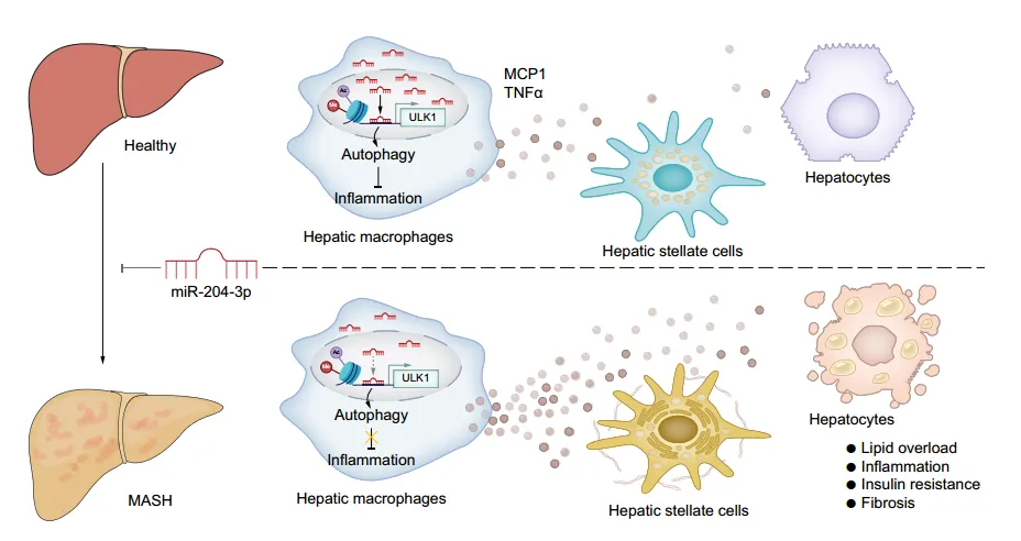
Increasing evidence suggests that innate immunity, especially macrophage activation, significantly promotes the development of metabolic dysfunction-associated steatotic liver disease (MASLD). Therefore, macrophage involvement in inflammation and interaction with hepatocytes and hematopoietic stem cells are potential therapeutic targets for MASLD. MicroRNAs (miRNAs) are a class of small non-coding RNAs that can bind to and degrade target mRNA. Abnormal miRNA expression regulates downstream genes crucial for the occurrence or progression of MASLD. In addition to post-transcriptional regulation of target genes, there is evidence that miRNAs exist in the nucleus of cells and play a regulatory role. Previous literature has reported that nuclear miR-204-3p in macrophages inhibits CD36 transcription and the formation of foam cell derived from macrophages, highlighting the nuclear function of miR-204-3p in regulating gene transcription, which warrants further investigation. Furthermore, miR-204-3p has anti-inflammatory effects in several inflammation-related disease models, but its significance in MASLD remains unclear.
Abstract
Recently, the team led by Sijia Liang from Sun Yat-sen University published a research paper titled “Nuclear miR-204-3p mitigates metabolic dysfunction-associated steatotic liver disease in mice” in Journal of Hepatology. This study revealed that miR-204-3p inhibits macrophage inflammation, coordinates macrophage interactions with hepatocytes and HSCs, and improves fatty liver disease. Macrophage miR-204-3p may be a therapeutic target for MASLD. In this study, TLR4 gene knockout THP-1 cells constructed by Ubigene were used to verify whether the anti-inflammatory effects of miR-204-3p depend on the inhibition of the TLR4/JNK signaling pathway.
Results & Discussion
Quantitative reverse transcription PCR shows that miR-204-3p expression decreased in the liver and macrophages of high-fat diet (HFD) mice and ob/ob mice. Small RNA sequencing analysis of samples obtained from participants shows that miR-204-3p levels were also downregulated in liver tissue and peripheral blood mononuclear cells (PBMCs) of MASLD patients, and miR-204-3p levels in PBMCs were negatively correlated with the severity of liver disease and damage. In summary, these results suggest that macrophage miR-204-3p levels are associated with hepatic steatosis, inflammation, liver injury, and disease occurrence.

Fig. 1. Reduced miR-204-3p levels in MASLD samples correlate with disease severity.
Considering the significant decrease in macrophage miR-204-3p levels during the progression of hepatic steatosis, the potential impact on fatty liver disease was studied. By injecting adeno-associated viruses overexpressing miR-204-3p (AAV-204-3p) into HFD mice, it was found that overexpression of miR-204-3p in macrophages alleviated hepatic steatosis, inflammation, and insulin resistance in mice, improving liver-related indicators. Similar results were obtained in methionine-choline-deficient (MCD) diet-induced MASH mouse models. Bone marrow transplantation experiments further confirmed the negative regulatory role of macrophage miR-204-3p in fatty liver disease. Overall, macrophage miR-204-3p improves experimental fatty liver disease in mice.

Fig. 2. Macrophage miR-204-3p ameliorates experimental steatohepatitis in mice.
The authors established a co-culture system of hepatic macrophages with primary hepatocytes or hematopoietic stem cells. Co-culture experiments showed that co-culturing with macrophages from HFD mice increased lipid deposition, inflammation, and insulin resistance in hepatocytes, and increased fibrotic gene expression in HSCs; whereas co-culturing with macrophages overexpressing miR-204-3p attenuated these changes. Knockdown of miR-204-3p enhanced JNK phosphorylation and secretion of MCP1 and TNFα in macrophages, but not in Tlr4-/-THP-1 cells, indicating that TLR4/JNK signaling inhibition is necessary for the anti-inflammatory effects of miR-204-3p. In macrophages overexpressing miR-204-3p, TLR4 expression and JNK phosphorylation were decreased, and secretion of inflammatory factors was reduced, thereby inhibiting hepatocyte steatosis and HSC activation. These results indicate that macrophage miR-204-3p reduces the secretion of pro-inflammatory cytokines, inhibiting hepatocyte lipid accumulation and HSC fibrotic activation.

Fig. 3. Macrophage miR-204-3p attenuates hepatocyte lipid accumulation and inflammation.
To elucidate the molecular mechanism of the anti-inflammatory effect of miR-204-3p in macrophages, the authors performed RNA sequencing on macrophages with upregulated miR-204-3p. They found that differentially expressed genes enriched in epigenetic modification-related gene ontology terms, with downregulated genes highly related to MASLD pathways. Fluorescence in situ hybridization revealed significant nuclear localization of miR-204-3p in liver macrophages, with reduced nuclear miR-204-3p levels in the liver of MASH and HFD-fed mice. The authors identified metabolism-related miR-204-3p regulatory genes and potential target genes ULK1 and NFXL1, and confirmed that ULK1 expression continuously increases after miR-204-3p overexpression. RNA hybridization prediction showed that miR-204-3p upregulates ULK1 promoter activity and transcript levels by binding to a conserved site at -153bp of the ULK1 transcription start site. Additionally, miR-204-3p promotes ULK1 promoter enrichment of RNAPII and H3K27 acetylation and H3K4 trimethylation in HMDMs, thereby enhancing ULK1 transcription.

Fig. 4. miR-204-3p acts in the nucleus and activates ULK1 transcription.
Given the important role of ULK1 in the initiation of autophagy, the authors investigated whether miR-204-3p restricts macrophage inflammation in an autophagy-dependent manner. They found that inhibiting autophagy reversed the suppression of the inflammatory response by miR-204-3p, while inducing autophagy enhanced the inactivation of the TLR4/JNK signaling pathway, indicating that miR-204-3p is associated with autophagy. Through experiments, the authors confirmed that miR-204-3p promotes autophagosome formation and fusion with lysosomes, increasing autophagic flux. Additionally, overexpression of miR-204-3p upregulated the expression of 15 autophagy-related genes, with a significant increase in the mRNA levels of ATG4A, AMBRA1, and ULK1. Furthermore, miR-204-3p regulates the initiation of autophagy through ULK1-dependent activation of the VPS34 complex, and inhibition of ULK1 eliminated the increase in Beclin1 phosphorylation and VPS34 activity.

Fig. 5. miR-204-3p inhibits macrophage inflammation by restoring ULK1-dependent autophagy.
To assess the functional relationship between miR-204-3p and ULK1 in hepatic steatosis, the authors injected AAV-control or AAV-204-3p into HFD-fed Ulk1-/- mice. They found that Ulk1 deletion led to increased liver weight/body weight ratios and that macrophage-specific miR-204-3p upregulation could not reverse this condition. Additionally, Ulk1-/- mice had decreased PparamRNA levels and increased levels of pro-inflammatory factors and inflammatory signaling molecules in the liver. These results indicate that the protective effect of miR-204-3p on liver fat accumulation is primarily dependent on ULK1.

Fig. 6. Ulk1 deficiency abolishes the protective effects of miR-204-3p on MASLD.
Conclusion
This study reveals a novel function of macrophage miR-204-3p, which acts through the transcriptional regulation of ULK1 expression. It enhances autophagic flux, reduces inflammation, and thereby limits fatty liver disease. Consequently, miR-204-3p and potential specific ULK1 agonists could serve as new therapeutic targets for metabolic dysfunction-associated fatty liver disease (MASLD).


 Back To Top
Our Solution Specialists will contact you as soon as possible!