Published on: July 24, 2024
Views:
--
Why does obesity increase the risk of asthma: TXNIP is the key!

Background
Obesity is associated with an increased incidence of asthma, but the specific mechanism by which obesity
triggers asthma is not fully understood. Previous studies have shown that obesity can lead to increased levels of
adipose factors and reactive oxygen species (ROS), affecting lung function and triggering airway inflammation. In
addition, Thioredoxin-Interacting Protein (TXNIP) plays a role in oxidative stress and inflammation, but its
specific role in obesity-induced asthma requires further investigation.
Abstract
Recently, Ji-Soo Jeong et al. published a research paper
entitled “The absence of thioredoxin-interacting protein in alveolar cells exacerbates asthma during obesity” in Redox Biology. The study reveals the role of Thioredoxin-Interacting Protein (TXNIP) in obesity-induced
asthma, providing a new perspective for the treatment of obesity-related asthma. The study utilized a TXNIP gene overexpression vector constructed by Ubigene to establish a TXNIP stable expressing A549 cell line, and investigated the effect of
TXNIP on inflammatory response by measuring levels of pro-inflammatory cytokines and ROS in the cells.
Construction of Obesity Mouse Asthma Model
Mice were fed with normal diet (ND) and high-fat diet (HFD) respectively, followed by intranasal
administration of Aspergillus proteinase (Asp) to induce asthma, and the symptoms were evaluated. Compared to ND
mice, HFD mice showed significantly increased Penh values at 20 and 40 mg/mL acetylcholine concentrations,
significantly increased eosinophil counts in bronchoalveolar lavage fluid (BALF), increased inflammatory cell
infiltration, increased mucus secretion, elevated serum total IgE levels, and increased levels of inflammatory
cytokines IL-4, IL-5, IL-13, and IL-17A. Compared to ND mice, HFD mice showed elevated serum FFAs levels, increased
adipose factors (leptin/adiponectin) ratio, and elevated intracellular ROS levels in BALF cells.
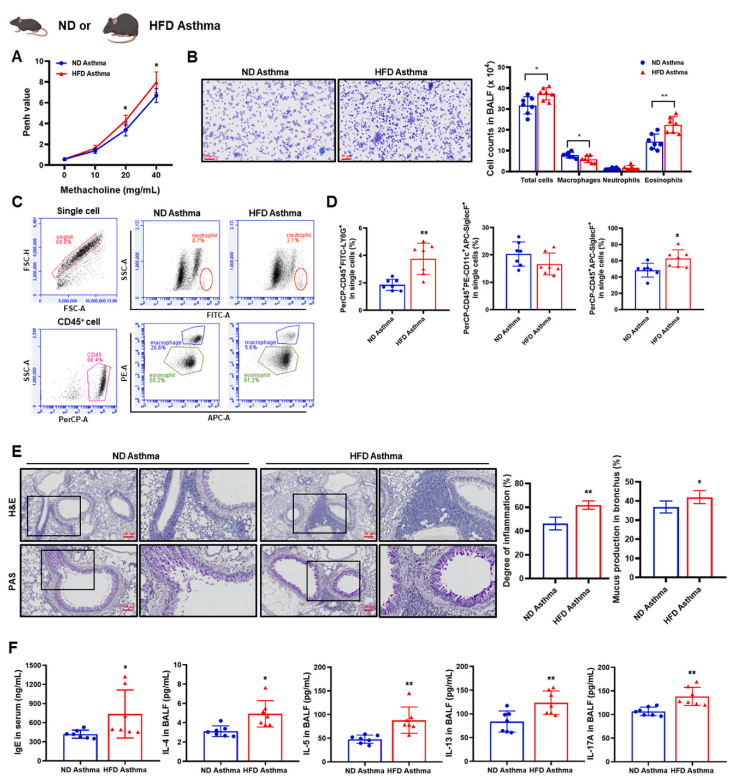
Figure 1 Comparison of asthma symptoms in mice fed with normal diet (ND) and
high-fat diet (HFD)
Construction of TXNIP Gene Knockout Mouse Model
TXNIPCre (TXNIP deficient in alveolar type II
cells) mice were used to validate the role of TXNIP in asthma induction. Protein expression of TXNIP and NLRP3 was
significantly increased in asthma patient samples compared to healthy controls; TXNIP and NLRP3 expression was
increased in alveolar type II cells of HFD mice compared to ND mice; TXNIPCre mice showed aggravated asthma symptoms
compared to TXNIPfl/fl mice, with increased eosinophil counts in BALF, increased inflammatory cell infiltration,
elevated serum total IgE and inflammatory cytokine levels, and increased expression of TXNIP and NLRP3
inflammasome-related proteins.
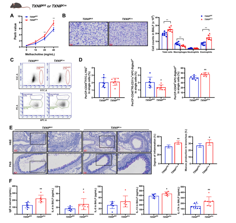
Figure 2 Using TXNIPCre mice to
validate the role of TXNIP in asthma induction
In vitro experiments
This study used the TXNIP gene overexpression vector constructed by Ubigene to establish a stable A549 cell
line expressing TXNIP. The effect of TXNIP on inflammation was studied through cell culture, analysis of mRNA
expression levels, and measurement of intracellular ROS levels. Compared to TXNIP-silenced cells, cells with TXNIP
overexpression showed decreased mRNA expression levels of pro-inflammatory cytokines IL-1β, IL-6, and TNF-α, as well
as decreased ROS levels.
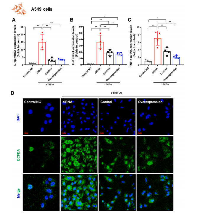
Figure 3 Expression of TXNIP affects levels of pro-inflammatory cytokines
and ROS in A549 cells.
Conclusion
The study results indicate that the increase in TXNIP levels in obesity-induced asthma is a compensatory
response to combat inflammation. This study provides new insights into the relationship between obesity and asthma,
particularly the role of TXNIP in regulating oxidative stress and inflammation, offering potential targets for
future treatment strategies.
ReferencesJeong, Ji-Soo, et al. "The Absence of Thioredoxin-Interacting Protein in Alveolar Cells Exacerbates Asthma during
Obesity." Redox Biology, vol. 73, 2024, 103193.
 Subscribe Us
 Gene Editing Services
 EZ-editor™
 Red Cotton Gene knockout Project


